$89 New patient Exams & X-rays / $35 Emergency Visits
Advanced Technology for Precise Care
At Grand Central Dentistry, we believe in providing the most accurate and comprehensive dental care to our patients. Utilizing cutting-edge technology, we enhance the precision of diagnoses and improve treatment outcomes. From high-definition intraoral cameras to 3D printing, our modern tools help ensure you receive the best care possible.
Our advanced dental technology not only provides better insights into your oral health but also helps us deliver treatments that are faster, more comfortable, and personalized. Read below to learn more about the technologies we use to provide you with the highest quality dental care.

Intraoral Camera
Our Intraoral Camera captures detailed, high-definition images of your teeth and gums, allowing us to spot potential issues that are not visible during a routine examination. Whether it's tooth decay, cracks, gum disease, or defective fillings, the intraoral camera helps us detect issues early.
By displaying these images on a screen, you’ll be able to see exactly what we see, making it easier for you to understand your oral health and participate in your treatment decisions. The ability to visualize potential problems in real-time leads to a more informed decision-making process and a better treatment outcome.
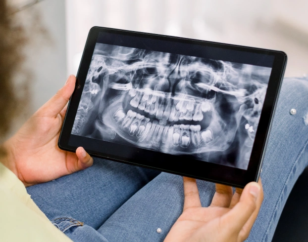
Digital Radiographs
Digital radiography is a revolutionary technology that replaces traditional X-ray films with advanced digital sensors. This results in clearer, high-definition images while using up to 80% less radiation. With these enhanced images, we can easily detect hidden cavities, bone infections, gum disease, abscesses, and even tumors that might be missed with a visual exam alone.
Our digital radiographs not only provide greater clarity but also promote faster diagnosis. They can be manipulated to enhance sharpness, contrast, and details, helping us make more accurate decisions. Plus, digital X-rays can be stored electronically for easy access and sharing with specialists, resulting in faster and more efficient treatment.
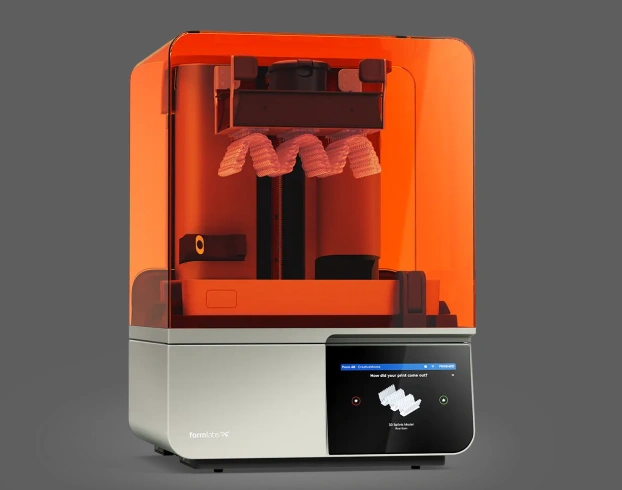
3D Printing
With 3D printing, we can create precise dental treatments faster and more affordably. From surgical guides for implants to temporary crowns and dentures, 3D printing allows us to offer a quicker, more personalized experience. Our in-house 3D printing capabilities save on lab fees, which means we can pass those savings onto you.
3D printing also allows you to preview your new smile before committing to permanent treatments. Whether you’re getting a crown or dentures, you can see how it will look in your mouth. If you want adjustments, we can make changes and print again until you’re completely satisfied with your result.
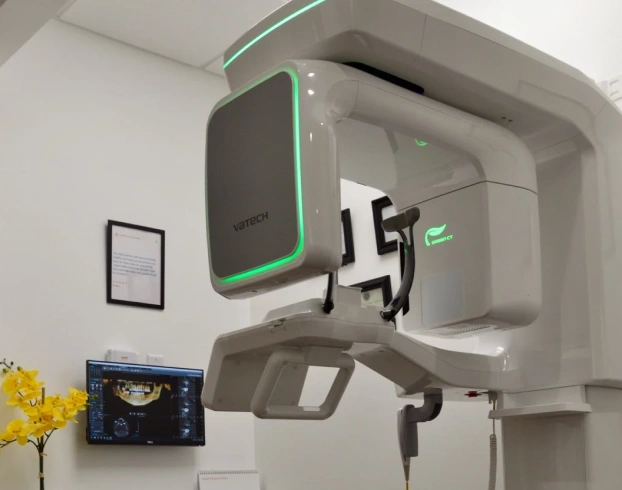
Cone Beam CT Scanner
Our Cone Beam CT (CBCT) scanner is an advanced imaging tool that provides detailed 3D scans of your jaws, teeth, and surrounding structures. This technology is especially beneficial for dental implant planning, as it helps us visualize the exact location for implant placement, improving accuracy and safety.
The CBCT scanner also helps identify infections, locate the source of pain, and visualize nerves during root canal procedures. In cases of sleep apnea, we can perform a comprehensive airway analysis to determine the best treatment.